Abstract
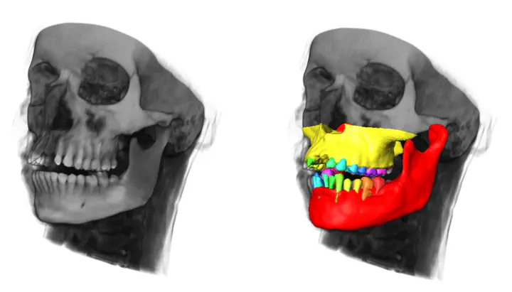
According to the American Academy of Implant Dentistry (AAID) the number of patients with bone-anchored dental implants is growing year by year. The goal is to reduce the costs of implant procedures and material, and at the same time ensuring implantation longevity, as well as patient safety and comfort. During surgery, the task is to place an implant into the bone in a stable manner and so that vital structures, such as dental roots, the inferior alveolar nerve, the sinus or major blood vessels are not damaged. One approach to achieve this goal are personalized surgical drill guides. These mechanical components are manufactured specifically for a given patient and can either be placed on top of the bone, gum or teeth. They must be designed accurately to obtain a stable implant position and to prevent the surgeon from damaging vital structures. A prerequisite for such a design is a pre-operative imaging of the relevant structures. Panoramic X-rays (orthopantomograms) are the predominant imaging technique in practice, but lack full three-dimensional (3D) information. 3D-imaging, however, becomes more and more accessible through the use of cone-beam CT data (CBCT or DVT). Hence, the task is to extract an accurate geometric representation of the relevant anatomical structures from such image data (segmentation). While CBCT is suitable for dental applications due to its small field of view and low dosage applied to the patient, it is less accurate than conventional CT and more prone to imaging defects such as noise and metal artifacts from dental fillings and brackets. This makes manual segmentation even more difficult and laborious.In this work, we present an automatic method for segmentation of all relevant structures of the jaw from CT data. This includes the mandibular and maxillary bone, the alveolar nerve and the overall detection and identification of existing (or missing) teeth. The only component missing in our work so far is an accurate segmentation of individual teeth (roots and crowns). Based on our tooth detection method, however, we are confident that this step can be reliably implemented in a future study
Hans Lamecker
Director, Software Development
Advancing 3D analysis, planning, design and manufacturing using innovative computational methods and tools